 |
||||
Silicic acid deposition in diatoms |
||||
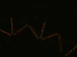
| Different kinds of dyes or stains, called probes, are used to examine the chemical processes and changes that occur in a phytoplankton cell. The images here show the same cells stained with the same probe and viewed with different light filters. The first set of images are Thalassionema cells. | ||||
|
|
|
||
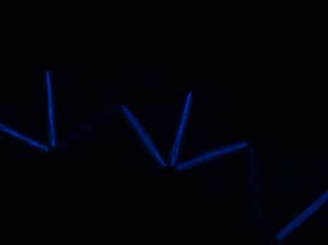
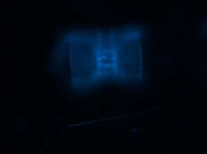
| The Thalassionema cells were collected from Monterey Bay and the probe was added to the water sample in the lab. After 24 hours, these images were taken. This image was taken with a chlorophyll filter, and the red spots show chloroplasts in the living cells. | These are the same cells, photographed with a D4 or DAPI filter set. The blue light emitted from the probe shows where silicic acid has been deposited over the 24 hour incubation period. |
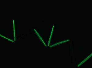
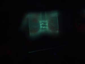
This image shows the silicic acid deposition under a different filter set, D5 or Long-pass DAPI. This experiment examines how silica deposition happens in diatoms in the ocean. |
||
|
 |
 |
||
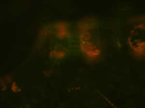
Chlorophyll filter, showing Stephanopyxis cells. These cells were incubated for 24 hours in the lab, as described above.
|
Stephanopyxis cells with D4 filter showing silicic acid deposition. |
|
||