 |
||||
Light microscopy |
||||
| Researchers have many tools in their arsenal for viewing and identifying phytoplankton. Initial viewing might take place with a field scope while still at the sampling site, but closer examination of the samples generally takes place in the lab with the help of a light microscope. A single scope may offer a variety of different magnifying lenses and light options. Four different viewing options are described below. By altering the path that the light takes through the scope to the viewer's eye, dramatically different images of the same phytoplankton cell can be created. | ||||
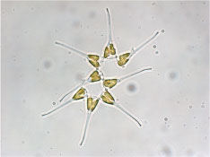
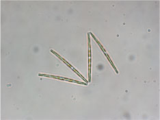
Brightfield Brightfield images use white light, and are illuminated from below. This is the simplest method of light microscopy, and highlights the natural colors present in a cell. |
|
|
||
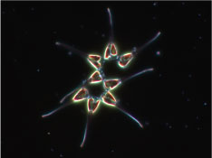
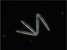
Darkfield Darkfield excludes light that is not scattered by the phytoplankton cell. As a result, only the cell itself is illumiated while the background remains dark, making cells "pop."
|
 - - |
|||
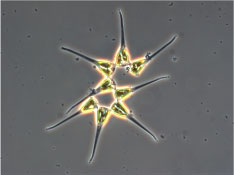
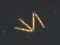
Phase Contrast Phase contrast enhances the contrast of clear or colorless objects. A glass plate inserted into the optical path enhances the phase shift of light passing through transparent material, creating a glowing ring around the image. |
 - - |
|||
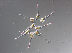
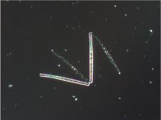
Differential Interference Contrast Also known as Nomarski Interference Contrast, DIC uses a complex lighting scheme that interprets the optical density of transparent material. The results are similar to phase contrast, but without the glowing ring effect.
|
 - - |
|||
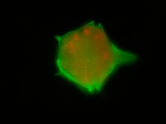
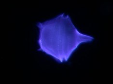
Epifluoresence In epifluorescence microscopy, a beam of light causes a cell to fluoresce. The emitted light is then focused by the objective lens. Stains are commonly used for highlighting specific proteins or molecules of interest. The images at right are stained with calcofluor, which highlights the cellulose plates in dinoflagellates. Different filters produce the two images. |
  |
|||
